

Pathology labs across Boston are dealing with more slides, more cases, and less room to slow down. The city has one of the highest concentrations of academic medical centers, teaching hospitals, and biotech affiliated research labs in the country, which means pathologists here are often reviewing complex cases under tight turnaround expectations from referring doctors and researchers. Many of these labs have already moved from physical glass slides to digital scans, a shift usually called digital pathology. The next step many are exploring is computer vision: software that can look at those digital slide images and help flag, measure, and organize what matters most, before a pathologist even opens the case. This is not about replacing a pathologist's judgment. It's about giving them better information, faster, so their expertise goes further.
This piece walks through what computer vision digital pathology actually looks like in practice, how it fits into a lab's existing setup, what compliance actually requires, and how a Boston lab might realistically start.
What Computer Vision Actually Does in a Digital Pathology Workflow
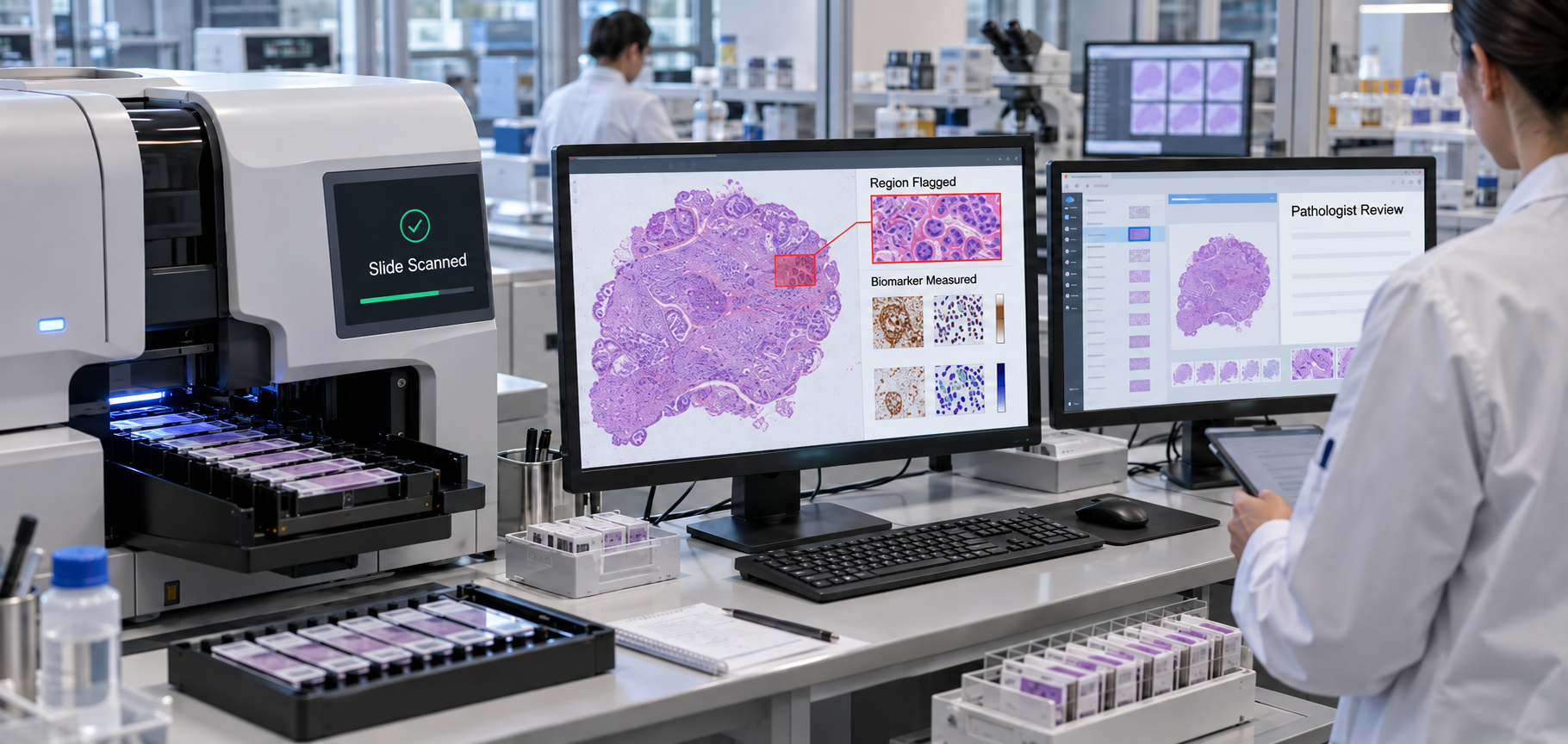
Here's the basic flow. A tissue sample on a slide gets scanned into a high resolution digital image. This is called whole slide imaging, or WSI for short. Instead of a pathologist immediately putting that image under review, computer vision software can scan it first. It looks for specific things: regions that look abnormal, areas with unusually high cell activity, or patterns that match known disease markers.
For example, in a biopsy being checked for cancer, the software might use tumor detection deep learning to highlight the regions most likely to contain tumor tissue, count how many cells are actively dividing, or measure the strength of a specific biomarker stain, a process pathologists call IHC scoring, where IHC stands for immunohistochemistry, a staining technique used to detect specific proteins in tissue. The software doesn't decide what the diagnosis is. It organizes and quantifies the visual information so the pathologist can review the case with that context already in front of them, instead of scanning the whole slide manually from scratch. This is a form of AI assisted histopathology that many labs are beginning to pilot.
In short, how does AI analyze a pathology slide? It scans the digital slide image, identifies and measures specific patterns like abnormal cell regions or biomarker levels, and presents those findings to the pathologist as a starting point for their review, not as a final answer.
Where This Fits into a Boston Lab's Existing Workflow
One of the biggest hesitations pathologists have about adopting new technology is workflow disruption. Nobody wants a tool that means rebuilding how the lab operates. The good news is that computer vision doesn't usually require that. Most Boston area labs, especially those connected to academic medical centers and teaching hospitals, already run whole slide imaging scanners as part of their digital pathology workflow automation setup. Computer vision typically sits on top of that existing infrastructure as an added layer, rather than a replacement for it.
In practice, this means the software connects to the lab's existing systems. Most labs run something called a Laboratory Information System, or LIS, which tracks cases, samples, and results, so this kind of LIS integration AI pathology work means the computer vision output shows up as part of the case the pathologist is already reviewing, not in a separate tool they have to check on the side.
We've built similar workflow automation layers before, including an AI powered clinical workflow platform designed for secure, structured review processes in healthcare settings, so we understand that the technical layer only works if it fits cleanly into how the people using it already work. If you want to see how this kind of technology is actually built, our AI computer vision services team works on exactly this: models that detect, measure, and organize visual data for real operational use, not just research demos.
Compliance, Validation, and Trust: What Boston Labs Need Before Adopting AI
This is the part that matters most, and it's worth being direct about it. Computer vision tools in pathology are decision support, not autonomous diagnosis. They assist a pathologist's review. They do not replace it, and no responsible vendor should claim otherwise.
Before a Boston lab adopts any AI tool for slide review, a few things need to be true. First, the lab's accrediting body, the College of American Pathologists, has published guidance on how labs should validate whole slide imaging and AI tools before using them clinically, meaning the lab needs to test the tool against its own cases and confirm it performs reliably before relying on it. This kind of HIPAA compliant digital pathology setup also needs to follow HIPAA, the federal law governing patient privacy, since slide images are tied to real patient records. Labs also operate under CLIA, the federal standards that govern clinical laboratory testing quality in the U.S., so any new tool needs to fit within those existing quality requirements, not work around them.
On the regulatory side, some computer vision tools for pathology have received FDA clearance, but that clearance is almost always for a specific, defined use, like flagging a certain type of tissue abnormality, not a blanket approval for the whole technology. It's worth checking a vendor's FDA clearance status for the exact use case you're considering, rather than assuming FDA cleared applies broadly. The College of American Pathologists' own guideline on validating whole slide imaging for diagnostic purposes is a good starting point for labs wanting to understand what that validation process actually involves.
Why Boston Diagnostic Labs Specifically Benefit
Boston's pathology labs handle a genuinely high volume of complex cases. Between academic medical centers, teaching hospitals, and labs affiliated with the city's dense biotech and research sector, the caseload tends to be both larger and more specialized than in many other regions. Referring oncologists and researchers often expect fast, well documented turnaround, especially for cancer cases where treatment decisions are waiting on the result.
This is exactly the kind of environment where AI diagnostic lab automation helps most, not by replacing careful review, but by helping labs triage: flagging which cases look most urgent, so a pathologist's time goes to the right slide first, and by handling repetitive counting and measurement tasks that eat up time without needing a pathologist's full clinical judgment. If you want to see how this fits into the broader picture of healthcare focused technology work, that's something we cover in our healthcare AI solutions as well.
Getting Started: A Practical Adoption Path
Labs that adopt this kind of technology successfully tend to start small rather than trying to overhaul everything at once. A realistic path looks like this:
- Pick one use case first, for example biomarker quantification for a single cancer type, rather than trying to cover every kind of case at once.
- Run it alongside your pathologists, not instead of them. Compare the software's output to what your pathologists find manually, and use that comparison to build trust in the tool before relying on it more.
- Expand gradually. Once one use case is validated and your team is comfortable with it, add additional case types or workflow steps.
This slower, staged approach is also what most pathology image analysis software validation processes expect, so it tends to align naturally with the requirements covered above. If your lab is exploring what a partner for this kind of work looks like, our team provides AI development services in Boston built around exactly this kind of staged, workflow first approach.
Frequently Asked Questions
Does computer vision replace pathologists in diagnosis?
No. It flags, measures, and organizes visual information for the pathologist to review. The final diagnosis stays with the pathologist.
Is AI based digital pathology FDA approved?
Some computer vision tools for pathology have FDA clearance, but it's typically for a specific, narrow use rather than the technology as a whole. It's worth confirming clearance for the exact use case you need.
How long does it take to integrate computer vision into an existing digital pathology setup?
It varies by lab, but starting with a single use case as a pilot is generally faster than trying to roll out full coverage across every case type at once.
What's the difference between digital pathology and AI powered digital pathology?Digital pathology refers to the scanning and viewing infrastructure, turning glass slides into digital images. Computer vision, or AI, is an added layer on top of that infrastructure that analyzes those images.
Bringing It Together
Computer vision isn't a replacement for the pathologists who do this work every day. It's a way to help their time and expertise go further, especially in a city like Boston where case volume and complexity are both high. Labs that start with one clear use case, validate it carefully against their own compliance requirements, and expand gradually tend to see the most reliable results.
If your lab is exploring what this could look like for your workflow, feel free to reach out to the team at Theta Technolabs at sales@thetatechnolabs.com. We're happy to talk through what a realistic first step would be for your specific setup.


_Enhancing%20Driver%20Safety%20and%20Compliance%20with%20Web%20Apps%20in%20the%20Logistics%20Sector_Q3_24.jpg)


_Integrating%20Chatbots%20Into%20Your%20Application.jpg)
_How%20much%20does%20it%20cost%20to%20create%20an%20android%20app%20in%202024%20for%20Startups_%20A%20detailed%20guide_Q2_24.jpg)
_Key%20Trends%20in%20Healthcare%20Software%20Development%20for%20the%20Future_Q2_24.jpg)
_Best%20iOS%20App%20Development%20Company_%20Enhancing%20User%20Engagement%20with%20Push%20Notifications_Q2_24.jpg)
_Chatbots%20for%20Event%20Management%20and%20Hospitality%20Services_Q1_24.jpg)
_Choosing%20the%20Right%20App%20Development%20Company_%20A%20Comprehensive%20Guide_Q1_24.jpg)