Dental diagnosis depends heavily on imaging. Every treatment decision — from a simple filling to a complex root canal or periodontal intervention — begins with what the dentist sees in an X-ray, panoramic scan, or CBCT image. In New York's competitive private dental market, where patient volumes are high and appointment windows are tight, the pressure to read imaging accurately and efficiently is constant.
AI-powered dental imaging analysis can support that process. Computer vision systems trained on dental imaging data can flag findings for dentist review, highlight regions of interest, and help clinicians focus attention on areas that warrant closer examination. The goal is not to replace the dentist's clinical judgment. It is to give dentists a more consistent, structured layer of imaging support that can contribute to improved AI imaging diagnostic accuracy in dental clinics across New York.
AI imaging systems support dentists during image review but do not replace clinical judgment.
The Diagnostic Challenge in Dental Imaging
Manual imaging review in busy dental clinics creates real operational challenges that compound over time:
- Early-stage finding detection is genuinely difficult: Subtle interproximal caries, early bone loss, and hairline root fractures are clinically documented as challenging to detect consistently on manual X-ray review, particularly under time pressure. This is where AI caries detection in dental imaging can provide meaningful supplementary support for dentist review.
- Clinician variability in interpretation: The same radiograph reviewed by two experienced dentists can yield different interpretations. This variability is a documented clinical reality, not a criticism — and it is one area where structured AI imaging analysis can contribute to greater consistency across a practice.
- Time pressure reduces review depth: A dentist seeing thirty patients in a day reviews imaging under very different conditions than one seeing ten. High-volume New York dental clinics feel this pressure acutely, and computer vision dental imaging tools can help maintain review consistency regardless of daily patient load.
- Patient communication gaps: When findings are subtle or complex, explaining them to patients without visual annotation is difficult. AI-generated highlighted imaging reports can support clearer patient communication and informed consent conversations.
According to clinical research, interproximal caries miss rates in manual radiographic review can be significant — particularly in busy practice environments where imaging is reviewed under time pressure.
How AI Imaging Can Support Dental Diagnostics
Computer vision systems designed for dental imaging analyze X-rays, panoramic images, and CBCT scans by identifying visual patterns associated with specific clinical findings. The system processes image data and highlights regions where patterns statistically associated with caries, bone loss, periodontal conditions, root anomalies, or lesions are detected — presenting these findings to the dentist as flagged areas with associated confidence levels.
The dentist reviews every flagged region and applies clinical judgment to determine whether a finding is clinically significant, requires further investigation, or can be monitored. The AI does not diagnose. It does not generate treatment recommendations. It surfaces image regions that the dentist should examine closely, structured in a way that supports faster and more consistent review.
To understand what this looks like in a real clinic day — consider a dentist reviewing bitewing X-rays for a patient presenting with mild sensitivity. Under normal review conditions, a subtle interproximal lesion at the early enamel stage may not be visually prominent enough to trigger immediate concern. An AI imaging system analyzing the same X-ray flags the region with a confidence score, drawing the dentist's attention to an area that warrants a closer look. The dentist examines it, applies clinical judgment, and decides whether to monitor or intervene. The AI did not make the diagnosis. It made sure the dentist looked at the right place. That is the specific value AI imaging support delivers in a high-volume New York dental practice
It is equally important to note that no AI imaging system detects all findings. False negatives — cases where a clinically significant finding is present but not flagged by the system; are a documented limitation of all current AI imaging tools. The dentist's own clinical assessment and full patient history remain the primary basis for diagnosis at all times.
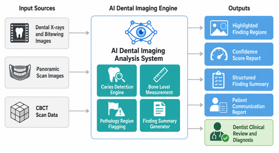
Figure: AI Dental Imaging Analysis Workflow in Dental Clinics
Some peer-reviewed clinical studies have reported improvements in early-stage finding detection rates when AI imaging support was used alongside dentist review — though study designs, imaging types, and outcome measures vary widely, and results from one clinical environment do not reliably predict outcomes in another. Well-implemented AI dental diagnostic tools in New York clinics can serve as a structured second layer of review that complements the dentist's own assessment rather than replacing it.
For dental clinics exploring broader AI-driven healthcare services, dental imaging analysis is one component of a wider AI integration strategy that can extend across patient management, treatment planning support, and clinical documentation.
Key Capabilities AI Can Bring to Dental Imaging
When implemented for dental clinic workflows, AI imaging systems can support several specific diagnostic functions:
- Caries detection and region highlighting: The system flags regions across bitewing and periapical X-rays where imaging patterns are consistent with carious lesions at various stages, presenting highlighted areas for dentist review.
- Bone level measurement support: AI can assist in measuring crestal bone levels across panoramic and periapical images, providing structured data that supports the dentist's periodontal assessment process.
- Root and canal morphology flagging: For complex restorative and endodontic cases, computer vision for dental X-ray analysis in dental clinics can flag unusual root curvature, canal configurations, or calcification patterns that may affect treatment planning.
- Pathology region highlighting: Lesions, periapical abscesses, cysts, and other radiolucent or radiopaque anomalies can be flagged for dentist attention and clinical assessment.
- Structured finding summaries: AI-generated imaging summaries can support patient communication, clinical documentation, and AI dental treatment planning support workflows by organizing findings in a structured, reviewable format.
Integration and Workflow Considerations
AI dental imaging systems are typically designed to integrate with existing practice workflows through DICOM compatibility, allowing them to connect with the imaging equipment and practice management systems dental clinics already use. Computer vision oral diagnostics platforms generally operate as a layer that processes images as they are captured, presenting flagged findings alongside the original image for dentist review at the chair or in a back-office review workflow.
Implementation requires configuration to the clinic's specific imaging equipment, validation of system performance on the clinic's own imaging data, and staff training on how to interpret and act on AI-generated findings. Computer vision dental imaging in New York clinics should be introduced as a clinical support tool with clear protocols for how dentists engage with flagged findings — not as a standalone diagnostic system.
Compliance, Clinical Oversight, and Patient Trust
AI dental imaging tools used in clinical environments in the United States may be subject to FDA regulatory oversight depending on their intended use and the claims made about their outputs. Dental clinics evaluating these systems should confirm the regulatory status of any tool under consideration with their legal and compliance teams before deployment.
Patient imaging data handled by AI systems must meet HIPAA requirements. Any system accessing, processing, or storing patient dental records must be designed with appropriate data encryption, access controls, and audit logging from the outset.
AI imaging diagnostic accuracy tools in dental clinics are clinical support systems. All diagnoses, treatment recommendations, and patient care decisions remain the responsibility of the licensed dentist. Transparency with patients about AI use in their care is both an ethical expectation and a trust-building opportunity for New York dental practices that choose to communicate it clearly.
Why New York Dental Clinics Are Evaluating This Now
New York's dental market is one of the most competitive in the United States. Private practices, group dental organizations, and specialty clinics operate in an environment where patient expectations for thorough, technology-supported care are high and growing.
In 2026, AI dental imaging tools are available at the clinic level — with several platforms already deployed in dental practices globally — though adoption in individual clinics varies significantly based on integration readiness, budget, and clinical workflow design. For New York dental clinic owners and dentists asking how AI can improve diagnostic accuracy in their practice, the technology is no longer a future consideration. It is a present-tense infrastructure decision that early-adopting practices are beginning to evaluate and implement.
Conclusion
AI imaging can meaningfully support diagnostic accuracy in New York dental clinics by providing a structured, consistent layer of imaging analysis that complements the dentist's clinical review. The technology flags findings, highlights regions of interest, and helps clinicians direct attention more efficiently — without generating diagnoses or replacing the judgment that only a licensed dentist can provide.
Implementation requires the right integration approach, HIPAA-compliant data handling, and a clear clinical protocol for how AI findings are reviewed and acted upon. The dentist remains the authority at every step. To explore how a purpose-built system can be designed for your clinic, connect with an experienced AI development company New York that understands both the clinical environment and the regulatory requirements.
Frequently Asked Questions
1. What does AI dental imaging diagnostics mean in clinical practice?
It refers to computer vision systems that analyze dental X-rays, panoramic images, and CBCT scans to flag findings for dentist review. Understanding how AI can improve diagnostic accuracy in New York dental clinics starts with recognizing that these systems highlight regions of interest for the dentist to assess — they do not generate independent diagnoses or treatment recommendations.
2. Does AI replace the dentist in making diagnoses?
No. AI imaging systems in dental clinics are clinical support tools. They flag regions in imaging data that warrant closer examination. All diagnoses, clinical interpretations, and treatment decisions are made by the licensed dentist who reviews the AI-flagged findings alongside their own assessment and the patient's full clinical history.
3. How is patient data privacy handled in AI dental imaging systems?
Any AI system that accesses or processes patient dental imaging data must be designed to meet HIPAA requirements. This includes encrypted data transmission and storage, role-based access controls, and audit logging of all data interactions. Dental clinics should confirm HIPAA alignment with their legal team before deploying any AI imaging system.
4. What imaging types can AI dental systems analyze?
Most AI dental imaging platforms are designed to analyze bitewing X-rays, periapical X-rays, and panoramic images. Some systems also support CBCT scan analysis. AI caries detection in dental imaging is most commonly applied to bitewing and periapical dental X-ray analysis, where early interproximal caries detection is clinically most valuable.
Build AI Imaging Capabilities for Your Dental Practice
Theta Technolabs builds custom AI and computer vision solutions for healthcare organizations across web, mobile, and cloud platforms. If your dental clinic in New York is evaluating AI-powered imaging diagnostics, reach out at sales@thetatechnolabs.com to discuss your requirements.


.png)
.png)


.png)

.png)
.png)
.png)


.png)
.png)
.png)
.png)

.png)


.png)


_How%20Cloud%20Solutions%20Are%20Enhancing%20Remote%20Patient%20Monitoring%20in%20Healthcare_Q4_25.jpg)
_Streamlining%20Appointment%20Scheduling%20with%20Cloud%20Computing%20in%20Dallas%20Healthcare_Q4_25.jpg)
_The%20Impact%20of%20Cross-Platform%20Apps%20on%20Real%20Estate%20Market%20Trends%20in%20Dallas_Q3_24-1.jpg)
_How%20AI%20is%20Enhancing%20Construction%20Site%20Surveillance%20and%20Security%20in%20Dallas_Q3_24-1.jpg)
_Web%20Apps%20for%20Retail%20and%20eCommerce_%20Streamlining%20Operations%20and%20Reducing%20Costs_Q3_24.jpg)
_Enhancing%20Driver%20Safety%20and%20Compliance%20with%20Web%20Apps%20in%20the%20Logistics%20Sector_Q3_24.jpg)


_Integrating%20Chatbots%20Into%20Your%20Application.jpg)
_How%20much%20does%20it%20cost%20to%20create%20an%20android%20app%20in%202024%20for%20Startups_%20A%20detailed%20guide_Q2_24.jpg)
_Key%20Trends%20in%20Healthcare%20Software%20Development%20for%20the%20Future_Q2_24.jpg)
_Best%20iOS%20App%20Development%20Company_%20Enhancing%20User%20Engagement%20with%20Push%20Notifications_Q2_24.jpg)
_Chatbots%20for%20Event%20Management%20and%20Hospitality%20Services_Q1_24.jpg)
_Choosing%20the%20Right%20App%20Development%20Company_%20A%20Comprehensive%20Guide_Q1_24.jpg)